Diffusion Magnetic Resonance Imaging
Project Summary
Diffusion Magnetic Resonance Imaging (DMRI) is a technique that produces in vivo images of biological tissues by exploiting the constrained diffusion properties of water molecules. DMRI can potentially be used to infer the organization of several structures in biological tissues. For instance, the extraction of neuronal fibers from brain DMRI can help understand brain connectivity in the corpus callosum, cingulum, thalamic radiations, optical nerves, etc. This has lead to numerous clinical studies showing how DMRI can be used to study brain development, multiple sclerosis, amyotrophic lateral sclerosis, stroke, Alzheimer's disease, schizophrenia, autism, and reading disability. In order to make DMRI beneficial in both diagnosis and clinical applications, it is of fundamental importance to develop algorithms for analyzing DMRI data. Researchers at CIS are developing methods for estimation, processing, segmentation and registration of diffusion tensor imaging (DTI) and high angular resolution imaging (HARDI).

The problems we are trying to solve are

  1. Registration using algebraic methods
  2. Segmentation using algebraic methods
Registration of diffusion tensor images
We consider the problem of registering diffusion tensor images under various local deformation models. Existing methods for DTI registration are computationally intensive and good initialization is critical. Our objective is to develop simple linear registration algorithms that can be used for initializing computationally intensive methods. In [1,2], we proposed an algebraic method for the registration of DTI data. We derive the so-called Diffusion Tensor Constancy Constraint, a generalization of the Brightness Constancy Constraint for 2D images to diffusion tensor data. Under the standard Euclidean metric in SPSD(3), we show that for various local deformation models, such as translational, rigid, and affine, together with the finite strain reorientation scheme as shown in Figure 1, the DTCC leads to a linear relationship between the parameters of the 
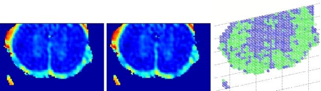
deformation, the DT data and its first order partial derivatives. Figure 2 shows the results of our registration algorithm using a multi-scale implementation.

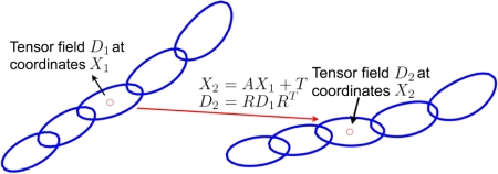
Figure 1: Registration of two DTI data sets under affine deformation model with finite strain reorientation scheme.


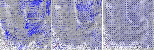
Figure 2: (Left and center) Deformation at scale 4 and 2. (Right) Final deformation.
Segmentation of diffusion tensor images
We aim to develop algorithms for separating a DT image into multiple regions. A region could be an actual anatomical structure, such as the corpus callosum in a brain image. Alternatively, a region could be a collection of fibers that have the same or similar function. In either case, the problem is to cluster a collection of tensors or fibers into groups according to some measure of similarity. We are studying metrics on diffusion tensors and fibers that are not only mathematically grounded but also biologically meaningful. Using these metrics, we are developing nonlinear dimensionality reduction techniques that will transform the DT data into a collection of clusters in a Euclidean space. The low-dimensional representation of the original DT data is then be segmented using standard clustering techniques. Figure 3 shows the results of our algorithm on segmenting the white and grey matter in the spinal cord.

Figure 3: (Left and center) apparent diffusion coefficient images for two slices of the spinal cord. (Right) fiber clustering results.

Work supported by startup funds from Johns Hopkins University, and by grants NSF CAREER IIS-04-47739.
Publications
[1]
A. Goh and R. Vidal.
European Conference on Computer Vision, pages 514-525, LNCS 3953, Springer , May 2006.
[2]
A. Goh and R. Vidal.
An Algebraic Solution to Rigid Registration of Diffusion Tensor Images.
IEEE International Symposium on Biomedical Imaging, pages 642-645, April 2006.